SINGLE CELL GEL ELECTROPHORESIS
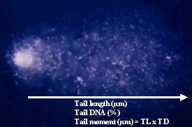
This technique has also been called "Comet assay" after the shape of the images (comets) seen under the microscope. It is a microgel electrophoresis technique which allows to measure DNA damage cell by cell. The alkaline version, introduced by Singh et al. in 1988, detects DNA breakage, alkali labile sites, open repair sites and cross links. For this technique cells are mixed with agarose gel, which is spread onto a microscope slide. The cells are lysed with high salt concentrations and detergents. The remaining nuclear DNA is then denaturated in an alkali buffer and electrophoresed in the same buffer. The DNA fragments migrate out of the nucleus, towards the positive pole. After electrophoresis, the slides are stained with a fluorochrome, like ethidium bromide. An image analysis system is used to measure several damage parameters. The most useful are tail length (TL), measured from the center of the nucleus towards the end of the tail, the percentage of DNA in the tail (TD) and tail moment (TM = TL x TD)
To
allow detection of oxidative DNA damage, the Fpg enzyme (Formamidopyrimidine DNA
glycosylase), which detects mainly 8-hydroxydeoxyguanine
and so called Fpg-sensitive sites, may be included (Hartwig et al., 1996).
Characteristics
of COMET test:
Advantages/ Disadvantages
|
Advantages |
Disadvantages |
|
|
Confounding factors:
controversial
References: